| Home |
| Acknowledgments |
| Conventions |
| Glossary |
| Maps |
| References |
| Links |
| Articles |
| Thumbnails |
| Species
list |
| Family |
| Next
species |
Additional Photos
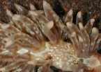
cerata detail

side
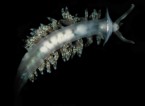
underside

light
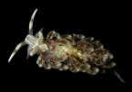
with yellow
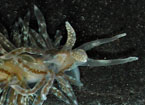
digestive gland detail

young, 3 mm

feeding
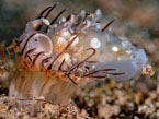
predation?
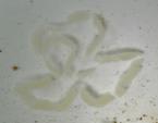
egg mass
_______________
GALLERY

Baeolidia salaamica (Rudman, 1982)
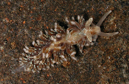
| Maximum size: 20 mm. Identification: This species is similar to Baeolidia moebii. However, it differs from that species in lacking extensive ramifications of the digestive gland in the notum, foot, rhinophores and cephalic tentacles (though it occasionally has them on the sides of its head). Its cerata are more numerous and slenderer than in B. moebii and they usually lack an obvious blue subapical band. Unlike in B. moebii, the cerata tips have prominent cnidosacs. Natural history: Baeolidia salaamica is a moderately common species. It's occasionally found in protected rocky habitats in as little at 1 m (3 ft). However, it is most common in Halimeda kanaloana beds at depths of 10-14 m (33-46 ft). It appears to be nocturnally active, concealing itself under rocks during the day. It feeds on the anemone Exaiptasia diaphana. (Note 1) Its egg mass is a cream or peach spiral composed of a "kinked" ribbon. (Note 2) The eggs hatch in 4-5 days in the laboratory. Distribution: Big Island, Maui, Oahu and Kauai: widely distributed in the Indo-Pacific. Taxonomic notes: The photos labeled Berghia major in Kay, 1979; Hoover, 1998 & 2006 and Bertch & Johnson, 1981 are actually of Baeolidia salaamica. It was deleted in the 5th printing of Hoover, 2006. Some sites list this species as Spurilla salaamica. Photo: CP: 12 mm; dark; found by PF; Maalaea Bay, Maui; Sept. 28, 2008. Observations and comments: Note 1: Kelly McCaffrey and Jenna Szerlag observed one attacking an Exaiptasia diaphana at Maalaea Bay in 2022. Jenna reported: "This was found by Kelly this morning at Kealia 24ft. He saw the approach and the initial attack/reaction. I watched it pull and tug on the anemone's tentacles a few times. They appeared to spring back into place as if unsuccessfully chewed off." After the initial contact, the tips of the inner tentacles ruptured and acontia were extruded and withdrawn through the ruptures. (see photo sequence) Note 2: Perhaps, the variation in egg mass color is due to diet (both were freshly laid when photographed)? |
| Thumbnails |
Species
list |
Family | Next species | Top |